
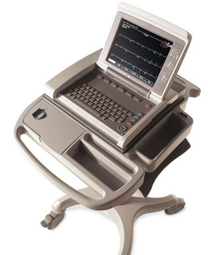
Evaluating Heart Health with Sound Waves An echocardiogram, also known as a cardiac ultrasound, is a non-invasive diagnostic tool that uses high-frequency sound waves to create detailed images of the heart. This advanced imaging technique allows our veterinary team to evaluate the structure and function of your pet's heart, providing valuable insights into their cardiovascular health.

Echocardiograms are particularly useful in diagnosing and monitoring conditions such as heart murmurs, arrhythmias, cardiomyopathies, and other heart-related disorders. This non-invasive imaging modality is safe, painless, and does not involve any radiation exposure.
Our facility is equipped with advanced echocardiography technology, ensuring accurate and detailed imaging of your pet's heart. Additionally, our skilled veterinary professionals are well-versed in interpreting the echocardiogram results, enabling us to provide prompt and effective treatment plans tailored to your pet's specific needs.