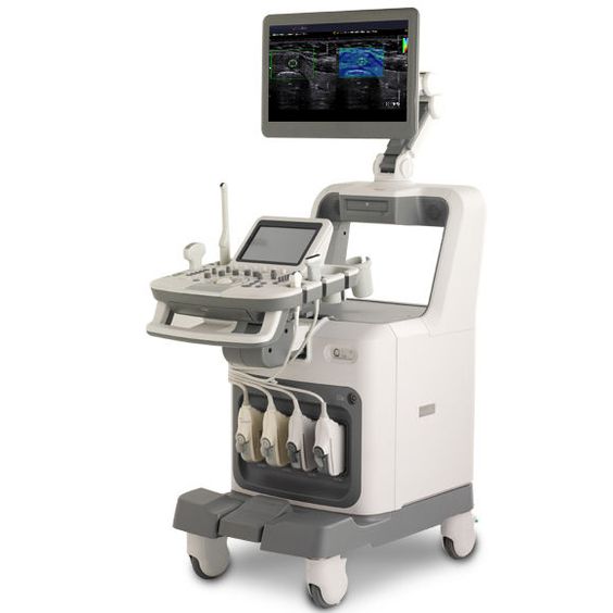
Unveiling Internal Insights with Sound Waves Ultrasonography is a diagnostic imaging technique that utilizes high-frequency sound waves to produce detailed images of a patient's internal organs. Unlike other imaging modalities like X-rays, this non-invasive method does not employ radiation. Instead, ultrasound waves are used to create visual representations of the structures within your pet's body.


Prior to the procedure, your pet should be fasted to prevent food in the stomach from obstructing the view of the organs and to facilitate sedation if needed.
If sedation is administered, our team will ensure your pet has fully recovered before allowing them to leave the clinic. Additionally, fine-needle aspirates of organs or lymph nodes can be performed under ultrasound guidance, depending on the size and location of the desired area.